A cataract is a clouding of the natural lens of the eye. The light entering the eye scatters and images can become blurry. When cataracts interfere with your daily activities then it is likely time to consider cataract surgery, the only effective treatment.
Cataract surgery
In cataract surgery, an ophthalmologist removes the cataract and replaces it with a clear artificial implant, restoring your vision. Routine cataract surgery is a painless 20-minute outpatient day procedure. For more information on what to expect with cataract surgery, including risks and benefits of the procedure, please visit the cataract surgery page.
Here we take you through the steps in cataract surgery with illustrations, video, and a photo-essay.
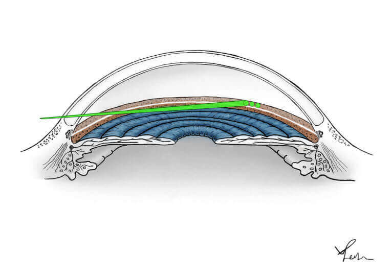
The cataract is removed through a 2-3mm incision with phacoemulsification, a specialized ultrasound technology
The support system is left in place to support a foldable one-piece IOL implant
Narrated video of routine cataract surgery
Step by step overview of cataract surgery
A 2.2mm self-sealing incision in the cornea allows the surgeon to access the cataract for removal
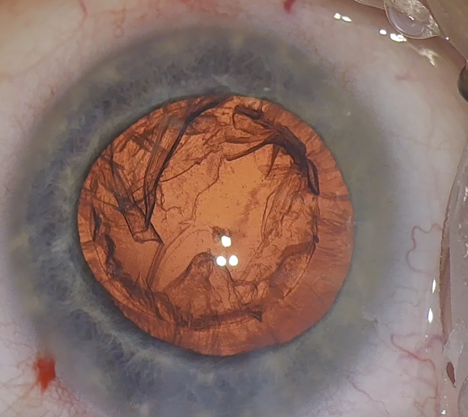
A capsulorhexis is a circular opening in the front of the capsule, the support system for the cataract. Here it is completed manually with forceps
The capsulorhexis opening allows the surgeon to remove the cataract without disturbing the support system, into which an artificial IOL implant will eventually be inserted
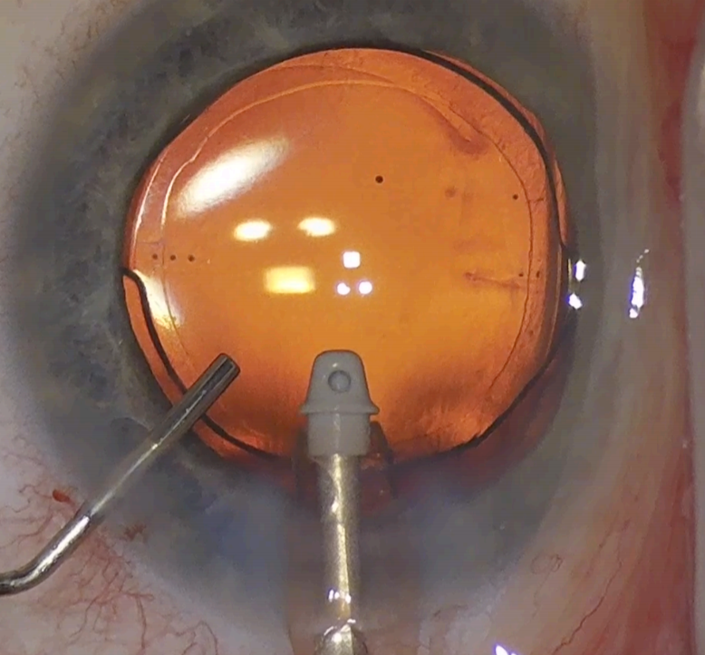
In hydrodissection, fluid is gently irritated between the capsule and the cataract, separating them and allowing the cataract to rotate so that it can be broken up into fragments
Phacoemulsification is a sophisticated ultrasound technology that allows the surgeon to break the cataract up into segments for removal. The surgeon is about to perform “the first crack” where the cataract is divided into two using a 'chopper' instrument
The cataract is then rotated and further divided into small fragments
The fragments are removed with a specialised fluid and the phacoemulsification ultrasound
The main part of the cataract, the nucleus, is now removed and the outer coat, the cortex, remains
The cortex is removed using a smaller hand-piece with specialized irrigation fluid and aspiration (I/A)
The capsule is then filled with a gel-like substance, viscoelastic, and an artificial IOL implant is folded and inserted into the eye through the 2.2mm incision
The implant unfolds and opens inside the capsule, the natural support system of the eye, that will keep it centred in place
The viscoelastic is removed. The IOL implant is nicely centered and the case is completed successfully
For information on cataracts and cataract surgery:
Wait times for cataract surgery are updated monthly, with wait 1 representing how long it takes in days to get in to see the surgeon in the office, and wait 2, how long it takes after that to get in for surgery.